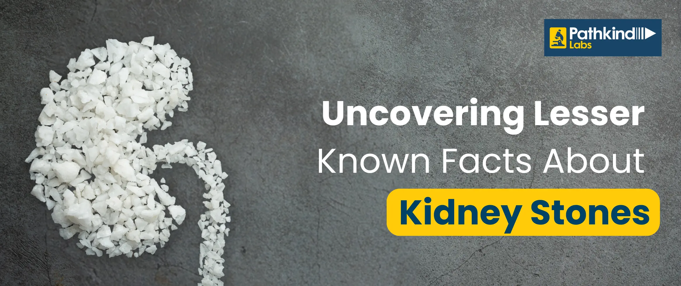
A kidney stone is a hard item formed from chemicals in the urine. The four forms of kidney stones are calcium oxalate, uric acid, struvite, and cystine. A kidney stone can be removed via shockwave lithotripsy, ureteroscopy, percutaneous nephrolithotomy, or nephrolithotripsy.
Urine contains many wastes. When urine becomes too concentrated, crystals develop. The crystals attract other elements and combine to form a solid that will continue to grow unless it is excreted in the urine. These substances are typically removed in the urine by the body's master chemist, the kidney. In most people, drinking enough liquid washes them out, or other substances in urine prevent stones from developing. Calcium oxalate, urate, cystine, xanthine, and phosphate are the chemical components that create stones.
After forming, the stone may remain in the kidney or migrate down the urinary tract to the ureter. Tiny stones can sometimes exit the body through the urine without causing significant pain. However, non-moving stones can induce a urine back-up in the kidney, ureter, bladder, or urethra, thus causing the pain.
A kidney stone usually does not produce symptoms until it moves around the kidney or enters one of the ureters. The ureters are tubes that link the kidneys to the bladder.
If a kidney stone becomes caught in the ureters, it can obstruct the urine flow, causing the kidney to enlarge and the ureter to spasm, which can be extremely painful. At that period, you may feel the following symptoms:
Other signs and symptoms may include:
You may be more likely to get kidney stones if:
A kidney stone is diagnosed using a medical history, physical examination, and imaging testing. Your doctors will want to know the specific size and shape of the kidney stones. This can be accomplished using a high-resolution CT scan from the kidneys to the bladder or a "KUB x-ray" (kidney-ureter-bladder x-ray), which will reveal the size and location of the stone. Surgeons frequently take the KUB x-ray to establish whether the stone is acceptable for shock wave treatment. The KUB test can monitor your stone before and after therapy, but a CT scan is often preferred for diagnosis.
In some cases, doctors will order an intravenous pyelogram, or lVP, an X-ray of the urinary system obtained after injecting a dye.
Second, your doctor will determine how to treat your stone. Blood and urine tests will be used to examine the health of your kidneys. Your overall health, as well as the size and position of your stone, will be considered.
Later, your doctor will determine the cause of the stone. After removing the stone from your body, your doctor will analyze your blood for calcium, phosphorus, and uric acid levels. Your doctor may also request you collect urine for 24 hours to test for calcium and uric acid.
Discover precision diagnostics with Pathkind Labs, a trusted name in diagnostic services in India. Our NABL accredited labs, equipped with advanced technology, are staffed by a certified team of over 200+ senior pathologists and 2000+ technicians. From tailored health check-ups to specialized tests in Oncology, Neurology, Gynaecology, Nephrology, and more, we've got your health covered. Skip the hassle with online booking for tests or check-ups, available for both lab visits and at-home blood collection. For a seamless experience and early detection, choose Pathkind Labs in Gurugram. Book your appointment today and experience diagnostics made easy.
© 2026 Pathkind Diagnostics Pvt. Ltd. All Rights Reserved | Unsubscribe